
Figure 1.
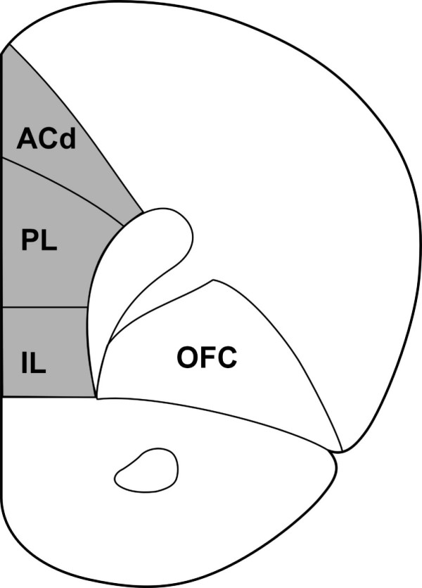
A coronal section through the rat brain illustrating the mPFC (shaded area). The mPFC consists of a dorsal mPFC (dorsal anterior cingulate cortex and dorsal part of the prelimbic cortex) and a ventral mPFC (ventral part of the prelimbic cortex and infralimbic cortex) [9]. Neurons were filled in coronal sections at approximately 1.7 mm to 3.7 mm from bregma. ACd: dorsal anterior cingulated cortex; PL: prelimbic cortex; IL: infralimbic cortex; OFC: orbitofrontal cortex. Modified after [35].