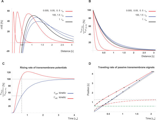
Figure 5. Time domain analysis of CIC system.
(A) V mE traces at different time points after starting to depolarize the synapse (V mP[x = 0] = 13 mV). Note that the VE pattern is already established at T = 0.005 (units of time-constant; τm = 48 ms) and that the VE-peak virtually reaches its final position and amplitude within 1.5 τm (72 ms). Each of the V mE traces is presented as percentage of the EPSP level at the position of the VE-peak (nVE), at the specific time point. The black trace depicts the V mE at time points of T = 1 whereas red and blue traces depict the V mE at time points lower or higher than T = 1, respectively. (B) For comparison, the conventional pattern of an EPSP along distance (V mP(X)) is plotted for the same time points as in A. The amplitude of each of the V mP traces is expressed as percentage of the potential at the signal's origin (synapse; X = 0). Color representation of the different time points is similar to (A). (C) The rising rate of V mE or V mP to steady-state level (red or blue, respectively) is simulated at the position of the VE-peak (X = 1.29). The amplitude is expressed as percent of the steady-state level at that position. Note that at the time V mP reaches 50% of its steady-state level, V mE has already reached its steady-state level and starts overshooting this after 0.6 τm (29 ms) V mE reaching peak of ∼20% above the steady-state levels at 1 τm (48 ms). Note that these kinetics lies within the duration of a synaptic current influx induced by a typical glutamatergic synapse (12–24 ms; see text for details). (D) The propagation pattern and rate of electrotonic signals along the internal cable (red; V mE) and along the external cable (blue; V mP) compared to the prediction of the conventional cable theory (gray; T = 2⋅λ). Following the conventional definition for electrotonic velocity we plotted, over time, the points where the transmembrane potentials reached 50% of its steady-state level (solid lines). Dashed red line depicts the rate the VE-peak approaches its steady-state position (dashed, horizontal gray line). At different positions along the internal cable, the potential develops toward positive or toward negative directions (as demonstrated in Figure 5A). For simplicity, only the region where V mE develops toward a positive direction was analyzed. This region lies above the dashed green line, which depicts the position where V mE = 0 at steady-state (X = 0.64 λ). Note the negligible difference between the predictions of the CIC model and the conventional model for the speed of electrotonic signals along the external cable (blue and gray lines, respectively).

