Figure 1.
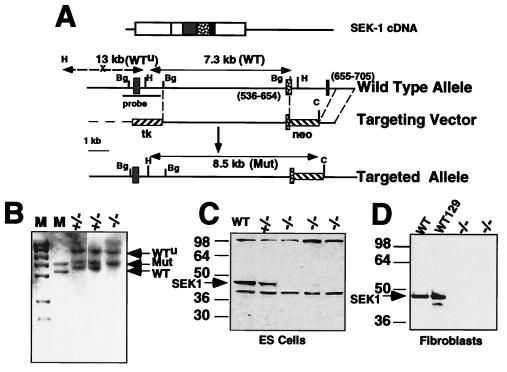
Targeted disruption of the SEK1 gene. (A) Targeting Strategy. The SEK1 cDNA is shown at the top; the coding region is represented by boxes, and the internal catalytic domain is represented by separate filled boxes corresponding to specific exons also shown on the genomic map. The structure of the wild-type and targeted alleles are shown. The SEK1 cDNA nucleotides representing the mutated exons are shown in parenthesis. Genomic DNA was analyzed for targeting events by Southern blot analysis by using the indicated probe and HpaI (H) and ClaI (C) digested DNA. The probe recognizes two wild-type fragments: a 13-kb 5′ (WTu) fragment whose structure is not affected by the SEK1 mutation and a 3′ 7.3-kb fragment (WT). An 8.5-kb fragment appears after homologous recombination with the targeting vector. (B) Identification of SEK1−/− ES cells. A Southern blot is shown of the parental SEK1+/− ES cell lines and an example of one SEK1−/− line derived by increasing the dose of G418. M represents markers. (C) Western blot analysis of SEK1 expression in targeted ES cells. Shown are SEK1 levels present in either the parental W9.5 ES cell line (WT) or one of the heterozygous targeted lines (+/−) and three independent SEK1−/− ES cell lines. No SEK1 protein was detected in any of the double knockout cell lines. (D) Western blot analysis of SEK1 expression in primary fibroblast cultures. No SEK1 was detected in any of the SEK1−/− fibroblast cultures.

