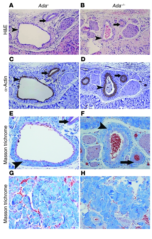
Figure 7. Ada–/– mice develop penile vascular damage and fibrosis subsequent to priapism.
(A–D) Histological examination of the vascular structures in the corpus spongiosum. (A and B) H&E staining. (C and D) Anti–α-SMA immunohistochemical staining. Arrowheads indicate intimal thickening with smooth muscle hypertrophy of the vascular wall in the deep dorsal vein; arrows indicate muscular hypertrophy of the arterial vascular wall. (E–H) Fibrosis in corpus spongiosum (E and F) and corpus cavernosum (G and H) visualized by Masson trichrome staining. Arrowheads denote the extensive fibrosis with extension into the intima of the deep dorsal vein; arrows indicate fibrosis around the lumen of the artery. Original magnification, ×100 (A–D); ×200 (E–H).

