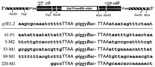
Figure 3.
Inverse PCR strategy to isolate and sequence the pB[Ccw] vector insertion site in transformant sublines. At the top is a schematic diagram (not to scale) of the vector insertion in the host plasmid showing the approximate location of the restriction sites and primers used for PCR. Forward (F) and reverse (R) primers are numbered according to their nucleotide position in piggyBac. The piggyBac sequence is shown shaded, the medfly white marker gene is open, and chromosomal sequence is hatched. Below are shown the piggyBac insertion site sequence in p3E1.2 and the proximal insertion site sequences from several of the transformant sublines.

