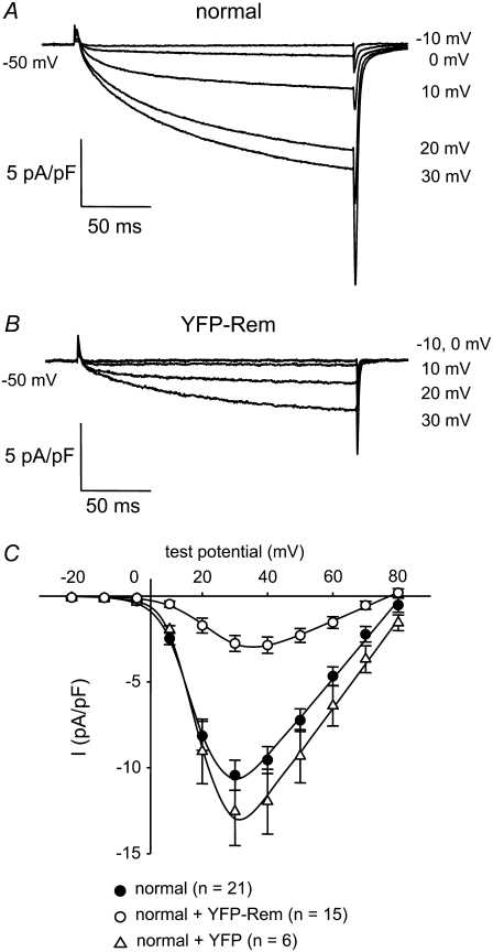
FIGURE 3.
YFP-Rem reduces skeletal muscle L-type Ca2+ current. Recordings of L-type Ca2+ currents elicited by 200-ms depolarizations from −50 mV to the indicated test potentials are shown for a normal, control myotube (A) or a normal myotube expressing YFP-Rem (B). (C) Comparison of I/V relationships for normal, control myotubes (•, n = 21), normal myotubes expressing YFP-Rem (○, n = 15), and normal myotubes expressing unconjugated YFP (Δ, n = 6). Currents were evoked at 0.1 Hz by test potentials ranging from −20 mV through +80 mV in 10-mV increments, following a prepulse protocol (28). The YFP-Rem I/V relationship represents pooled data from myotubes injected with 5, 10, 20, 50, and 100 ng/μl YFP-Rem cDNA; no significant difference in peak current density was found between these groups (p > 0.05, ANOVA). The smooth curves are plotted according to Eq. 2. The best fit parameters for each plot are presented in Table 2.

