FIGURE 1.
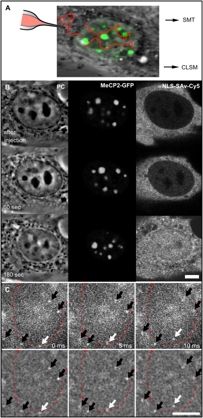
NLS-SAv-Cy5 complexes in living cells and their nuclear import. (A) Scheme of the experimental approach for confocal imaging and photobleaching experiments and for single-molecule detection. Mouse myoblast C2C12 cells transfected with plasmids coding for MeCP2-GFP (green) were microinjected into the cytoplasm with preformed NLS-SAv-Cy5 complexes as tracer molecules. Nuclear import was followed by confocal microscopy (CLSM), and after its completion, photobleaching experiments were performed. In a separate experimental setup, single molecules were tracked using a widefield fluorescence microscope (SMT). (B, left panel) Phase contrast (PC) images of a nucleus, time points of the image sequence as indicated. (B, middle panel) Green channel showing the distinct MeCP2-GFP labeling of the pericentric heterochromatin. (B, right panel) confocal time lapse images acquired in the red channel after the cytoplasmic microinjection of NLS-SAv-Cy5 complexes, demonstrating efficient nuclear import. (C) In the single-molecule setup, single SAv-Cy5 molecules could be observed while entering into the nucleus (white arrows) across the nuclear envelope (red dotted line). Further molecules could be identified and tracked within cytoplasm or nucleus (black arrows). In the upper panel unprocessed data are shown; in the lower panel, the effect of bandpass filtering used for further processing is demonstrated. Scale bars, 5 μm.

