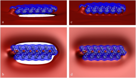
FIGURE 5.
Electrostatic potential isosurfaces for Lys13 adsorbed on a ternary (PC/PS/PIP2) lipid membrane. (a and b) Side and top views, respectively, of the system in the initial configuration. (c and d) Similar views for the final state of the system, after 500 ns. Lys13 van der Waals surfaces are colored in gray. The red surface represents Φ = −1.5 kBT/e (−37.5 mV) equipotential contour, and the blue mesh depicts the Φ = +1.5 kBT/e (+37.5 mV) equipotential contour. For clarity, the lipid membrane is not shown.

