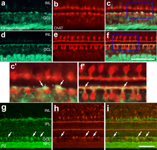
Figure 3.
Type-II cholinergic amacrine cells transiently express neurofilament during embryonic development. Vertical sections of chick retina labeled with antibodies to neurofilament (green) and ChAT (red). Sections were obtained from embryos at E8 (a-c) E11 (d-f), or from a postnatal chick (P2) (g-i). The boxed-out areas in panels c and f are enlarged 2.5-fold in panels c' and f'. Arrows indicate the dendrites of type-II cholinergic amacrine cells that co-localize immunoreactivities for neurofilament and ChAT. The calibration bar (50 μm) in panel f applies to panels a-f, and the bar in i applies to panels g-i. Abbreviations: ChAT – choline acetyltransferase; INL – inner nuclear layer; IPL – inner plexiform layer; GCL – ganglion cell layer; NFL – nerve fiber layer.

