Figure 3.

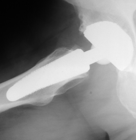
sagital radiograph of the Cut prosthesis. Figure 2 is showing the anteroposterior, figure 3 is showing the sagital radiograph 12 months after implantation of the implant in a 50 year old male patient.

sagital radiograph of the Cut prosthesis. Figure 2 is showing the anteroposterior, figure 3 is showing the sagital radiograph 12 months after implantation of the implant in a 50 year old male patient.