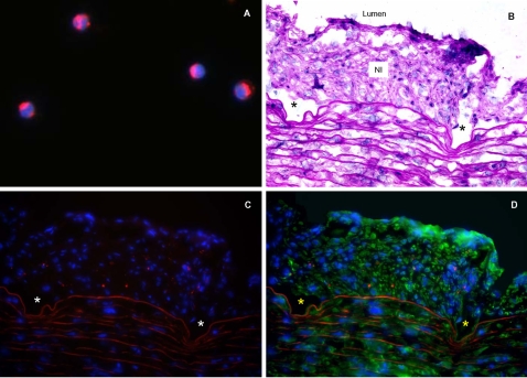
Figure 6. Differentiation of circulatory cells in vivo.
A: PBMCs labeled in vitro with PKH26 dye (red) and DAPI nuclear counterstain (blue); magnification: ×1000. These cells were injected into the carotid artery immediately after stent insertion. B: Cross section of native rabbit carotid artery 14 days post stent implantation (H&E stain,). C: PKH26-labelled PBMCs (red) were detected in the NI of stented artery. D: Some PKH26-labelled PBMCs (red) were also immunopositive for α-SMA (green). For panels B–D: arrows or autofluorescence delineate the internal elastic lamina; *denote sites of manually removed stent struts; magnification: ×400.

