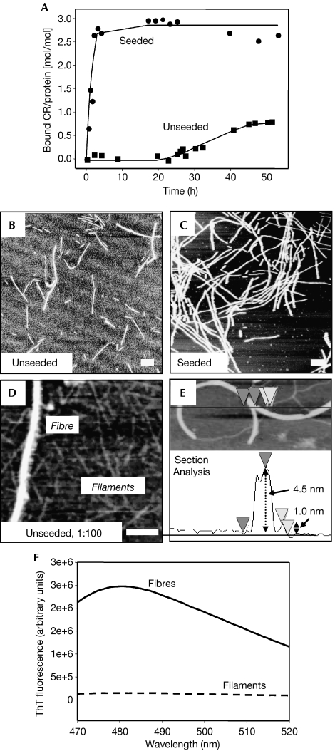
Figure 1.
Amyloid fibre assembly of NM monitored by congo red binding in quiescent conditions. (A) Unseeded (squares) and seeded with 4% w/w sonicated fibres (circles); 150 μg/ml soluble protein each. After completion of the assembly reaction, samples were investigated by AFM: (B) unseeded and (C) seeded; (D) reflects a diluted (1:100) sample of (B). (E) AFM section analysis to exemplarily determine filament and fibre heights. Scale bars, 200 nm. (F) ThT binding (excitation at 450 nm) of fibres and filaments. AFM, atomic force microscopy; CR, congo red; M, highly charged middle region; N, asparagine- and glutamine-rich region; ThT, thioflavin T.

