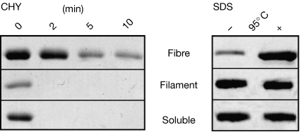
Figure 4.
Stability of filaments, fibres and soluble NM analysed by Coomassie-stained SDS–PAGE. Left panel: protease digestion (1/50 w/w chymotrypsin) at 37°C for 0, 2, 5 and 10 min. Right panel: SDS stability (2% w/v SDS) before (−) and after (+) incubation at 95°C for 10 min. The protein bands indicate monomeric NM. CHY, chymotrypsin; M, highly charged middle region; N, asparagine- and glutamine-rich region; SDS–PAGE, SDS–polyacrylamide gel electrophoresis.

