Figure 1.
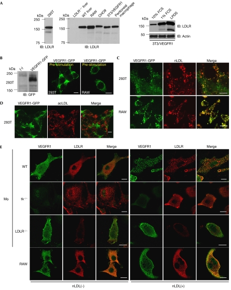
Native low-density lipoprotein induces endocytosis of VEGFR1. (A) Immunoblotting (IB) of whole-cell lysates (WCLs; 30 μg each) from human 293T cells (left) with human LDLR antibody and those from rodent cells with mouse LDLR antibody, including the liver of LDLR−/− and WT mice, RAW, CHO9, NIH3T3 cells overexpressing VEGFR1 (3T3/VEGFR1) and peritoneal macrophages (middle). Cell lines were cultured in 1% FCS for 24 h before protein extraction. WCLs from 3T3/VEGFR1 cultured in 10% and 1% serum, or LPDS were also subjected to anti-LDLR and anti-actin immunoblotting (right). (B) WCLs of 293T cells transfected with the VEGFR1-GFP expression vector or mock (−) were immunoblotted with GFP antibody (left). Membrane localization of VEGFR1-GFP transfected into 293T or RAW cells before nLDL stimulation (right). Scale bars, 10 μm. (C,D) 293T or RAW cells transfected with VEGFR1-GFP (green) were incubated with 10 μg/ml DiI-nLDL (red) (C) or 10 μg/ml DiI-acLDL (red) (D) for 30 min. Note the merged images with nLDL but not acLDL. Scale bars, 10 μm. (E) Peritoneal macrophages (MΦ) derived from WT, tk−/−, LDLR−/− and RAW cells were immunostained with VEGFR1 and LDLR antibodies before (−) and after (+) stimulation by nLDL at 100 μg/ml. acLDL, acetylated LDL; GFP, green fluorescent protein; LDL, low-density lipoprotein; LDLR, LDL receptor; LPDS, lipoprotein-deficient serum; nLDL, native LDL; VEGFR, vascular endothelial growth factor receptor; WT, wild type.

