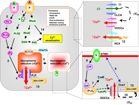
Figure 1.
A model for excitation/contraction coupling in a tonic smooth muscle cell in which sustained contraction involves Ca2+ entry through both VOCCs and SOCCs. (1) Physiologically, SOCE is initiated either by stimulation of receptors that couple through heterotrimeric GTP-binding protein (G proteins) to activate phospholipase Cβ (PLCβ) or (2) by stimulation of receptors that couple through tyrosine phosphorylation to activate PLCγ (Parekh and Penner, 1997; Patterson et al., 2002). This results in breakdown of phosphoinositide and production of IP3. (3) This second messenger activates IP3 receptors, which are ligand-gated Ca2+ channels located in the SR. The resulting release of Ca2+ into the cytoplasm causes a transient increase in [Ca2+]i, whereas (4) emptying of Ca2+ stores generates a retrograde signal that activates SOCCs in the plasma membrane, which are responsible for the sustained increase in [Ca2+]i after the initial Ca2+ transient. (5) Depleted Ca2+ stores generate a key messenger molecule called Ca2+ influx factor (CIF), which diffuses to the plasma membrane. (6) A cascade of plasma-membrane-delimited reactions in which (7) CIF displaces inhibitory CaM from the membrane-bound iPLA2, leading to iPLA2 activation and the generation of lysophospholipids (8) that in turn activate SOCCs. Ca2+ release from the SR causes the sensor (i.e., Ca2+-sensing STIM1 protein) to aggregate in areas close to the plasma membrane and to interact with SOCC (i.e., Orai1), which is believed to be the store-operated channel (9). On the other hand, (10) SOCC may also provide direct depolarization, independently of Ca2+ -activated Cl− channel VOCCs are opened by membrane depolarization due to initial Ca2+ -release from SR, which stimulates a Ca2+ -activated Cl− channel (11). (12) The plasma membrane NCX is involved in the regulation of Ca2+ homeostasis in blood vessels by contributing to Ca2+ entry. Finally, (13) NO/cGMP inhibits SOCCs, possibly by enhanced re-filling of the Ca2+ -stores. (14) Ca2+ sensitization might occur via agonist-induced activation of either the small G protein RhoA/Rho-associated kinase (Rho K) pathway or protein kinase C (PKC). A, agonist; CICR, Ca2+ -induced Ca2+ release; CIF, calcium influx factor; CLCA, Ca2+-activated Cl− channel; iPLA2, Ca2+-independent phospholipase A2; CaM, calmodulin; CPA, cyclopiazonic acid; DAG, diacylglycerol; G, GTP binding proteins; cGMP, guanosine-3′,5′-cyclicmonophosphate; IP3, inositol-1,4,5-trisphosphate; NCX, Na+/Ca2+ exchanger; NO, nitric oxide; PKC, protein kinase C; PLC, phospholipase C; R, receptor; S, putative Ca2+ sensor; SOCC, store-operated Ca2+ channel; STIM1, stromal-interacting molecule 1; TG, thapsigorgin; TK, tyrosine kinase; Vm, membrane potential; VOCC; voltage-operated Ca2+ channel.

