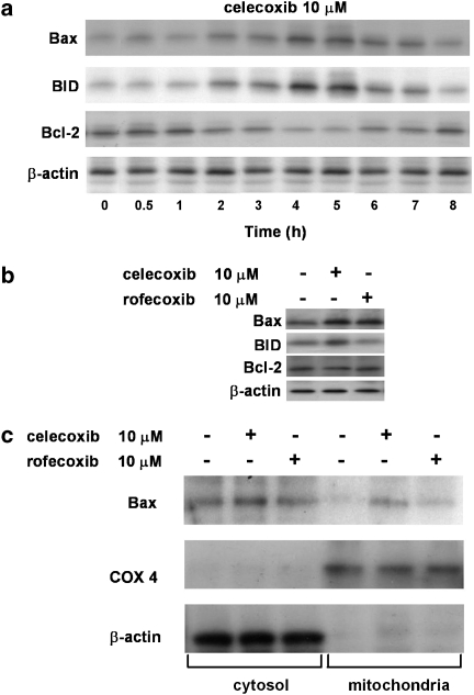
Figure 6.
Effect of celecoxib and rofecoxib on expressions of Bax, BID and Bcl-2. (a) HT29 cells were incubated with celecoxib (10 μM) from time 0 to 8 h. At the time indicated, cells were processed for western blot analysis as described in the Methods section. The immunoblot shown is representative of three separate experiments. (b) HT29 cells were treated with celecoxib or rofecoxib (10 μM) for 4 h and processed for western blot analysis as described in the Methods section. The immunoblot shown is representative of four separate experiments. (c) Immunoblot representing translocation of Bax to mitochondria in HT29 cells treated with celecoxib or rofecoxib (10 μM) for 4 h. COX 4 and β-actin are mitochondrial and cytosolic protein markers, respectively. The immunoblot shown is representative of three separate experiments.

