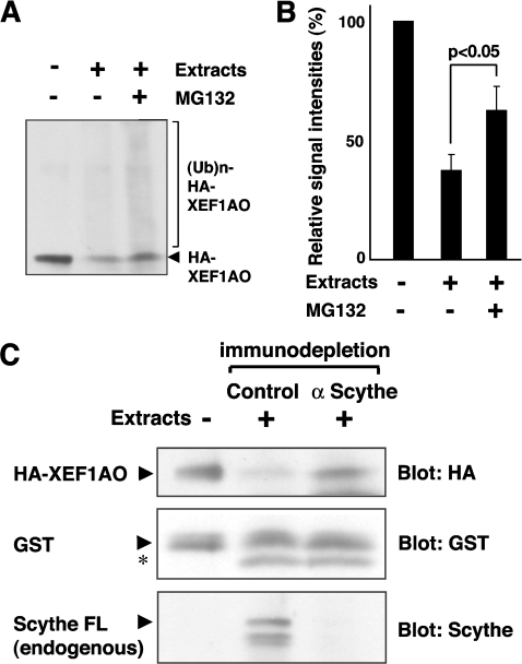
Figure 6. Scythe is essential for the proteasome-mediated degradation of XEF1AO in Xenopus egg extract.
(A) Bacterially expressed HA–XEF1AO was mixed with Xenopus egg extracts prepared from stage 18 embryos in the presence or absence of 100 μM MG132 as indicated. The mixtures were incubated at 37°C for 1 h and the reaction was stopped by the addition of SDS. The samples were immunoblotted using an anti-HA antibody to measure the amount of XEF1AO. (B) The intensity of the signals derived from XEF1AO and its polyubiquitinated forms in (A) was measured by Scion image and quantitatively represented. The results are shown as the means of duplicates for one out of three independent experiments. (C) Purified HA–XEF1AO was mixed with Xenopus egg extracts at stage 18 that had been immunodepleted either by anti-Scythe IgG or by control IgG. The mixtures were incubated at 25°C for 24 h, and the reaction was stopped by the addition of SDS, followed by immunoblotting with anti-HA antibody to measure the stability of HA–XEF1AO. As a control, bacterially produced GST was added in an equal amount to each sample and blotted with anti-GST antibody, and its stability was compared with that of HA–XEF1AO. The results of anti-Scythe immunoblotting confirmed appropriate immunodepletion of endogenous Scythe protein from the extracts. Arrows indicate the full-length form of corresponding proteins, and the asterisk (*) indicates a non-specific band. FL, full length.

