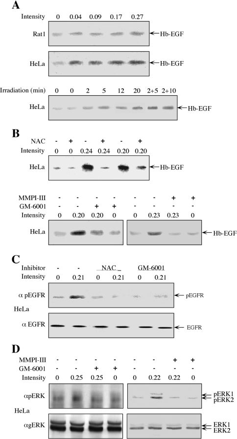
Figure 5. Involvement of Hb-EGF, MMPs and ROS in the irradiation-induced phosphorylation of ERKs.
(A) Serum-starved Rat1 (top panel) and HeLa (middle panel) cells were irradiated at 875 MHz with an intensity of 0.04, 0.09, 0.17 and 0.27 mW/cm2 for 10 min. For the time course determination, serum-starved HeLa cells were irradiated at 875 MHz with an intensity of 0.31 mW/cm2 for the indicated times (bottom panel). After stimulation, the starvation medium was collected, Hb-EGF was enriched using heparin beads (as described in the Experimental section) and subjected to Western blot analysis with anti-Hb-EGF antibody. (B) Serum-starved HeLa cells were incubated for 20 min with 2.5 mM NAC (upper panel), 0.5 μM GM-6001 (lower panel, left) or 0.4 μM MMPI-III (lower panel, right), or left untreated as a control. The cells were then irradiated at 875 MHz with the indicated intensities for 10 min. The release of Hb-EGF was detected as in (A). (C) Serum-starved HeLa cells were incubated with NAC (2.5 mM; 20 min), GM-6001 (0.5 μM; 20 min) or were left untreated. One plate from each treatment was irradiated at 875 MHz with an intensity of 0.21 mW/cm2 for 5 min, whereas the other plate was left untouched. The cells were harvested in RIPA buffer and subjected to Western blot analysis with anti-pEGFR (α pEGFR) or anti-EGFR (α EGFR) antibodies, as indicated. (D) Serum-starved HeLa cells were treated with GM-6001 (0.5 μM, 20 min), MMPI (0.4 μM, 20 min) or were left untreated. The cells were then irradiated at 875 MHz with an intensity of 0.25 or 0.22 mW/cm2, as indicated. The phosphorylation of ERKs was detected as described in the legend to Figure 1.

