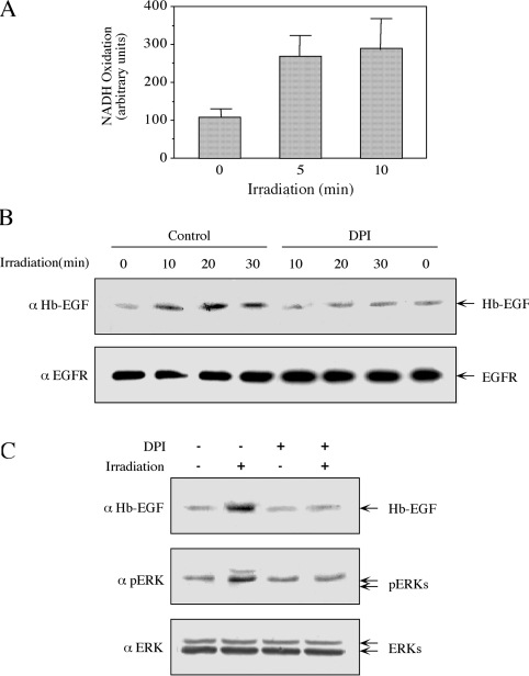
Figure 8. NADH oxidase is involved in the irradiation-induced release of Hb-EGF and phosphorylation of ERKs.
(A) Plasma membranes from serum-starved HeLa cells were isolated as described in the Experimental section. Membranes (5 μl of net membranes dissolved in 600 μl of buffer containing 250 μM NADH in PBS) were irradiated at 875 MHz with an intensity of 0.240 mW/cm2 for the indicated times. NADH oxidase activity was determined as described in the Experimental section. Results are means±S.E.M. for three independent experiments. (B) Plasma membranes of serum-starved HeLa cells were either incubated with 12 μm DPI (15 min) or left untreated, as indicated. The membranes were then irradiated at 875 MHz with an intensity of 0.200 mW/cm2 for the indicated times. The amount of Hb-EGF and EGFR was analysed by Western blotting with the indicated antibodies. (C) Serum-starved HeLa cells were incubated with 12 μM DPI for 30 min or left untreated as a control. Cells were then irradiated at 875 MHz with an intensity of 0.200 mW/cm2 for 10 min. The amount of Hb-EGF released and phosphorylation of ERKs was determined with the indicated antibodies, as described in the legends to Figures 1 and 5.

