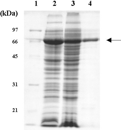
Figure 2. SDS/PAGE of GH5BG–thioredoxin fusion protein expressed in E. coli strain Origami B (DE3) after incubation in the presence of 0.5 mM IPTG at 20 °C for 12 h.
Lanes: 1, standard marker (Bio-Rad); 2, total protein of E. coli cells containing pET32a(+)/DEST-GH5BG; 3, soluble fraction of E. coli cells containing pET32a(+)/DEST-GH5BG; 4, purified thioredoxin–GH5BG. The arrow points to the thioredoxin–GH5BG.

