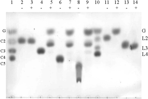
Figure 3. Hydrolysis of disaccharides and oligosaccharide substrates by thioredoxin–GH5BG detected by TLC.
The thioredoxin–GH5BG was incubated with 5 mM substrates for 30 min and the products were detected by the carbohydrate staining method described in the Experimental section. Samples were incubated with (+) or without (−) enzyme in 50 mM sodium acetate, pH 5.0, for 30 min at 37 °C prior to being spotted on silica-gel 60 F254 TLC plates, developed and charred with 10% H2SO4 in ethyl alcohol. Lanes: 1, glucose (G) and cello-oligosaccharides of DP 2–4 (C2–C4) marker; 2 and 3, cellobiose; 4 and 5, cellotriose; 6 and 7, cellotetraose; 8 and 9, cellopentaose; 10, standard laminari-oligosaccharides of DP 2–4 (L2–L4); 11 and 12, laminaribiose; 13 and 14, laminaritriose.

