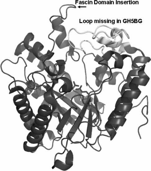
Figure 4. Active site of Candida albicans Exg structural model with differences in the loops around the active site found in rice GH5BG highlighted.
The 1CZ1 structure [9] is shown as a ribbon diagram coloured dark grey, with the loop after β-strand 7 of the (β/α)8 barrel shown in white and labelled to draw attention to its absence in rice GH5BG. The insertion of the fascin-like domain after the first helix of the extended loop after strand 1 of the β-barrel is indicated by the label. The catalytic acid/base (left) and catalytic nucleophile (right) are displayed in stick representation to indicate the location of the active site. The image was generated with Pymol [30].

