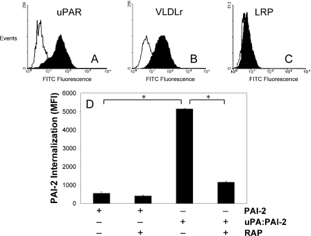
Figure 1. uPAR and VLDLr mediate the endocytosis of uPA–PAI-2 by MCF-7 cells.
MCF-7 cells were probed with 10 μg/ml of primary (A) uPAR, (B) VLDLr or (C) LRP polyclonal antibodies. These were detected using anti-rabbit IgG-FITC (1:50 dilution) and the cells analysed by flow cytometry, using propidium iodide to exclude non-viable cells. (D) MCF-7 cells were incubated in the presence or absence of RAP (200 nM) for 15 min at 37 °C, prior to analysis of PAI-2–Alexa Fluor®488 or uPA–PAI-2–Alexa Fluor® 488 internalization using the fluorescence quenching internalization assay (means±S.E.M., n=3; *P<0.05).

