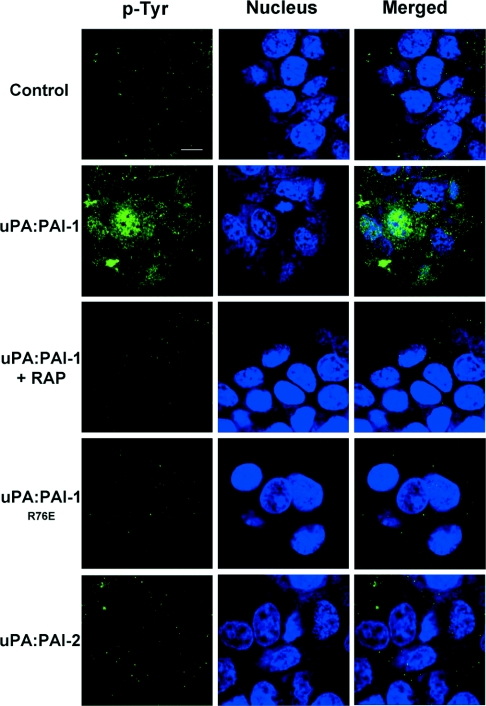
Figure 5. uPA–PAI-2 does not induce nuclear/cytoplasmic protein tyrosine phosphorylation.
MCF-7 cells were serum starved for 4 h and incubated in the presence or absence of RAP (200 nM) for 15 min at 37 °C, then incubated with 10 nM uPA, uPA–PAI-1, uPA–PAI-1R76E or uPA–PAI-2 for 30 min at 37 °C. The cells were washed with ice-cold PBS, fixed with 3.75% (w/v) paraformaldehyde and permeabilized with 0.2% Triton X-100. After incubation with 10 μg/ml anti-phosphotyrosine monoclonal antibody (PY20), the cells were washed and incubated with goat anti-mouse IgG-FITC (1:200 dilution) and TO-PRO 3 (1:400). After washing, the cells were analysed by confocal microscopy. The scale bar represents 10 μm.

