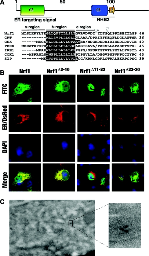
Figure 5. Nrf1 contains an ER-targeting signal sequence around the NHB1.
(A) NHB1 and its flanking residues are predicted to form a hydrophobic α-helix, whereas the residues around NHB2 may fold as a basic amphipathic α-helix and β-sheet structure. Multiple sequence alignments are shown of signal sequences from Nrf1 and other ER-resident proteins including CRT (calreticulin), CNX (calnexin), PERK (PKR-related ER kinase), IRE1 (inositol-requiring kinase 1), COX1 (cyclo-oxygenase 1) and S1P (Site-1 protease). (B) Expression constructs for wild-type Nrf1 and its specific deletion mutants around NHB1, along with an ER/DsRed construct encoding an ER marker protein, were cotransfected into COS-1 cells. Subsequent immunocytochemistry and imaging were performed as described in the legend of Figure 2. Scale bar=20 μm. (C) COS-1 cells were transfected with Nrf1/pcDNA3.1/V5His before being subjected to immuno-electron microscopy. The dots in original and enlarged images represent positive signals for ectopic Nrf1 protein.

