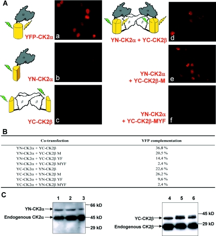
Figure 6. Visualization of interactions between CK2α and CK2β or CK2β mutants in living cells by BiFC.
(A) Immunofluorescence images of HeLa cells transfected with plasmids expressing the EYFP fragments fused to CK2α, CK2β or CK2β mutants as indicated in each panel. At 24 h after transfection, immunostaining of EYFP was performed to enhance the signal as described in the Experimental section. The schematic diagrams on the left of the images represent the experimental strategies used. (B) Quantification of the BiFC signal in cells co-transfected with different plasmid constructs. Results are expressed as the percentage of cells that were above the fluorescence threshold observed in cells expressing only YN-CK2α or YN-CK2β. (C) Western blot analysis of the levels of protein expression. Cells corresponding to panels d, e and f in (A) that expressed the indicated proteins were harvested, and the cell extracts were analysed by Western blotting using anti-CK2α (lanes 1–3) and CK2β (lanes 4–6) antibodies. Lanes 1 and 4, YN-CK2α+wild-type YC-CK2β; lanes 2 and 5, YN-CK2α+YC-CK2β-M; lanes 3 and 6, YN-CK2α+YC-CK2β-MYF. Molecular masses are indicated in kDa.

