Figure 3.
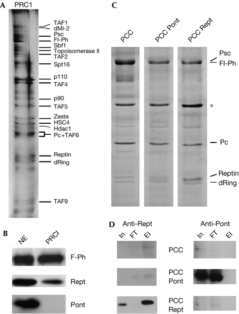
Rept is an integral component of PRC1. (A) SDS–PAGE (silver stain) of PRC1. Protein bands are labelled according to Saurin et al (2001). (B) Western analyses of PRC1. NE: 20 μg nuclear extract; PRC1: 15 μl PRC1. (C) SDS–PAGE (Coomassie blue) of reconstituted PCCs from Sf9 cells expressing Flag-Polyhomeotic (F-Ph), Psc, Pc, dRing (PCC), and Pont (PCC-Pont) or Rept (PCC-Rept). *Nonspecific Sf9 protein. (D) Western analyses of Rept (left) and Pont (right) profiles during purification of PCCs shown in (C). In: 1.5 μl (15 μg) Sf9 nuclear extract; FT: 1.5 μl anti-Flag affinity column flow through; El: 3 μl purified complex. PCC, PRC1 core complex; Pont, Pontin; Psc, Posterior sex combs; Rept, Reptin; SDS–PAGE, SDS–polyacrylamide gel electrophoresis.

