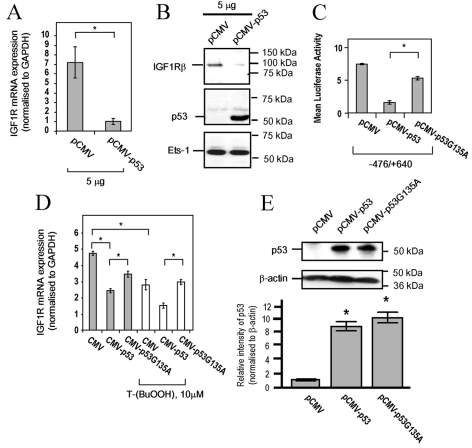
Figure 4. Repression of IGF1R by T-(BuOOH) is mediated by p53.
(A) IGF1R mRNA expression in control and p53-transfected WKY12-22 rat VSMCs, as determined by quantitative PCR. Results are means±S.E.M. (n=3). *P<0.05. (B) Western blot analysis of IGF1R, p53 and Ets-1 (loading control) in control and p53-transfected WKY12-22 rat VSMC lysates. (C) IGF1R promoter activity in WKY12-22 rat VSMCs co-transfected with −476/+640 IGF1R promoter–luciferase (1 μg/well in a six-well plate) and 0.5 μg/well of pCMV, pCMV-p53 or pCMV-p53G135A, as determined with the dual luciferase assay system. Results are means±S.E.M. (n=3). *P<0.05. (D) IGF1R mRNA expression in WKY12-22 rat VSMCs transfected with CMV, CMV–p53 or CMV–p53G135A in the presence or absence of 10 μM T-(BuOOH) for 24 h, as determined by quantitative PCR. Results are means±S.E.M. (n=3). *P<0.05. (E) p53 protein expression in WKY12-22 rat VSMCs transfected with 5 μg of pCMV, pCMV-p53 and pCMV-p53G135A. Lower panel shows p53 expression normalized to β-actin (arbitrary densitometric units). Results are means±S.E.M. (n=2). *P<0.05.

