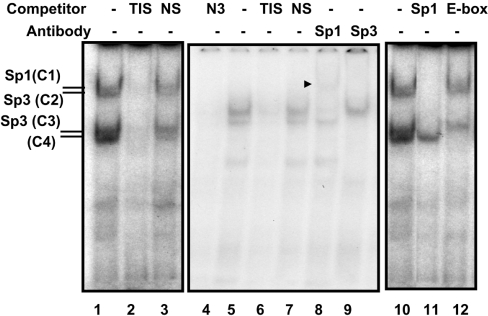
Figure 3. Sp1 and Sp3 bind to the NHE2 TIS.
Gel-shift analysis was carried out using 5 μg of C2BBe1 cell nuclear proteins. The end-labelled probe used in these assays was the double-stranded oligonucleotide from −12 to +20, shown as TIS in Table 2. Binding reactions were performed in the presence or absence of competitors (50-fold molar excess) or in the presence of anti-Sp1 and -Sp3 antibodies (2 μg/lane) as indicated at the top of the Figure. All oligonucleotides used in the experiments are shown in Table 2. Four bands showed specific binding to the −12/+20 probe and are indicated by C1–C4. To separate the DNA–protein complexes the gel represented by numbers 4–9 was run for a longer time than the others that resulted in weaker interactions of C4. NS, non-specific nucloetide; N3, an oligonucleotide from the NHE3 promoter that we have shown previously binds Sp1 and Sp3. An arrowhead indicates the Sp1-supershifted band.

