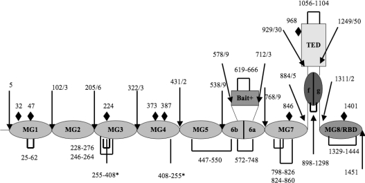
Figure 1. Schematic representation of the domain organization of the α2M monomer, based on alignment with the structure of C3 [17].
MG designates a macroglobulin domain; CUB, a domain found in a number of developmentally regulated proteins [32]; ‘bait’, bait region domain. Intramolecular disulfide bonds are shown as solid black rectangular loops. The two inter-chain disulfides (Cys255 and Cys408) [33,34] are shown as straight black lines and asterisked. The position of possible N-linked glycosylation sites are marked with black diamonds. Note that both MG6 and CUB are split domains whose primary structures are interrupted by other domains, the bait domain and TED respectively.

