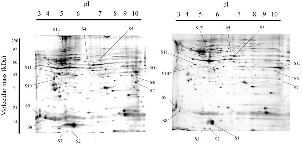
Figure 1.
Photographs of two-dimensional electrophoresis gels with annotation of the spots of identified proteins. The left image shows a silver-stained gel of embryo chick retina at ED7 and the right image is that of embryo chick retina at ED11. The proteins spots that increased or decreased in embryo days ED7 to ED11, and that were identified by PMF are shown. Spots S1 through S13 represent the annotated spots. The pI gradient of the first dimension electrophoresis is shown on the top of the gels, and the migration of molecular mass markers for SDS-PAGE in the second dimension is shown on the side of the gel. Representative gel images are shown.

