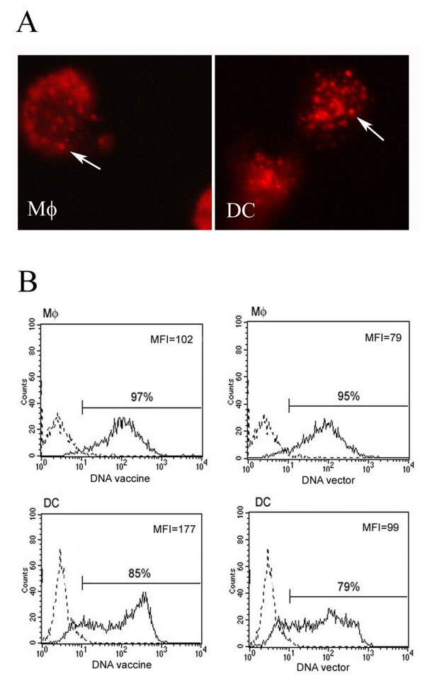
Figure 2.
Uptake of DNA-HSP65 by macrophages (Mφ) and DC. (A) Cells were stimulated for 4 h with Alexa Fluor 594-labeled DNA-HSP65 and analysed by fluorescence microscopy. Endocytic vesicles are indicated by white arrows. (B) Differential capacity of macrophages and DCs to uptake DNA vaccine. Cells were stimulated for 1 h with Alexa Fluor 488-labeled DNA-HSP65 and analysed by flow cytometry. These results are representative of three independent experiments. Black line: stimulated cells; dotted line: non-stimulated cells.

