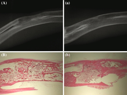
Fig. 4.
The radiograph of a experimental group rabbit at 9 weeks shows bone formation connecting both osteotomy sites. The defect is filled completely (A). The defect is filled completely with newly formed mature bone in H-E stain of Fig. 3A. (×6) (B). The radiograph of a control group rabbit at 9 weeks shows complete bone formation and remodelling in the defect (a). The defect is filled with newly formed mature bone in H-E stain of Fig. 3a (×6)(b)

