
Figure 6.
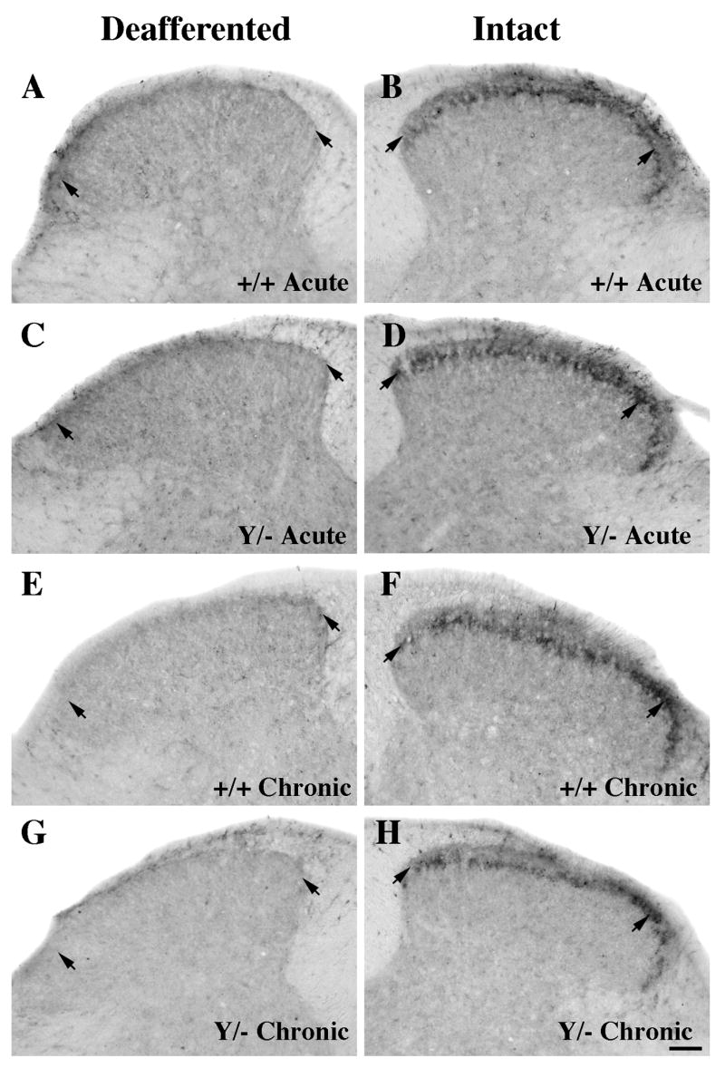
Comparison of P2X3 immunoreactivity at the center of the deafferented region (~T12-T13) following acute (A-D) and chronic (E-H) unilateral rhizotomy in wild-type (A-B, E-F) or L1 mutant (C-D, G-H) mice. The intact and deafferented dorsal horn images are from the same section, and all sections were processed together in the same experiment.
A–D: P2X3 expression on the intact side is concentrated in lamina II inner (short arrows) in both wild-type (B) and L1 mutant (D) mice. Five to seven days after rhizotomy, a large decrease is detected on the deafferented side in mice of both genotypes (A, C).
E–H: Three months post-rhizotomy, primary afferents labeled with P2X3 are still absent in both genotypes (E, G; short arrows). Scale bar A-H = 50 μm.