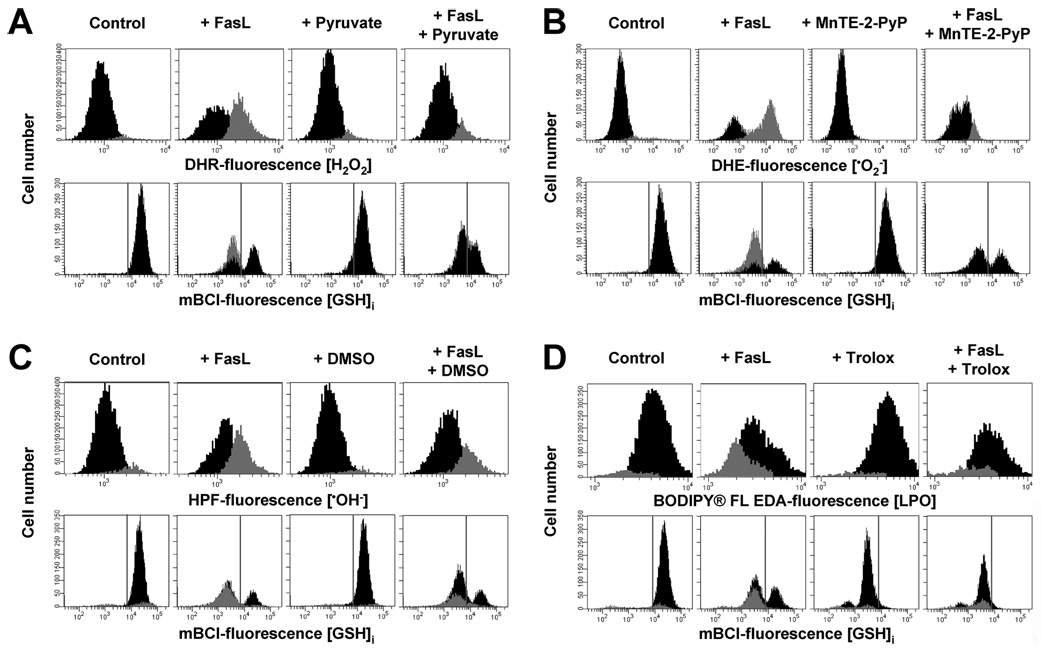
FIGURE 2. Reactive oxygen species scavenging by antioxidants does not affect FasL-induced GSH loss.
Reactive oxygen species formation and changes in GSHi were assessed by FACS. Apoptosis was induced in Jurkat cells by incubation with FasL (50 ng/ml FasL) for 4 h. The effect of the antioxidants 10 mm sodium pyruvate (for H2O2) (A), 2% Me2SO (•OH−) (B), 250 µm MnTE-2-PyP () (C), and 5 mm trolox (LPO) (D), was individually assessed. Jurkat cells were preincubated in RPMI for 1 h at 37 °C with the agents dissolved in either Me2SO or ethanol, and antioxidants remained throughout the experiment. In all cases, control conditions include vehicles at the same concentration. Populations were gated according to the differences in ROS levels. Data are expressed as frequency histograms of either changes in the fluorescence of the ROS-sensitive dyes (upper panels A–C) or mBCl (lower panels A–C). In A–C lower panels, GSH depletion is depicted as the appearance of a population of cells at the left of the gray solid lines. For mBCl fluorescence the solid line depicts the plots representative of n = 3 independent experiments.

