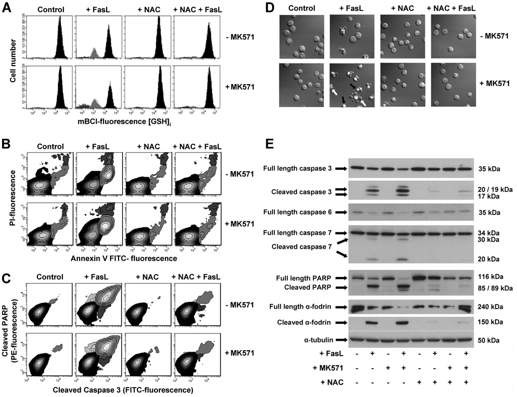
FIGURE 9. N-acetyl-l-cysteine protects against FasL-induced GSH depletion and apoptosis.
Apoptosis was induced in Jurkat cells by incubation with 25 (A) and 50 (B–E) ng/ml FasL for 4 h. MK571 (50 µm) and high extracellular NAC medium (+NAC) were added at the time of FasL stimuli. A, changes in GSHi were determined by FACS. Populations were gated according to their GSHi levels on an mBCl fluorescence versus forward scatter plot as explained under "Experimental Procedures." FasL-induced apoptosis was assessed by (B) phosphatidylserine externalization and loss of plasma membrane integrity; (C) simultaneous detection of cleaved caspase 3 and PARP; (D) DIC images depicting nuclear condensation, plasma membrane blebbing, and cellular fragmentation; and (E) immunoblot detection of full-length and cleaved forms of execution caspases 3, 6, and 7, as well as their substrates PARP and α-fodrin. Results are expressed as in Fig. 5. Plots, images, and blots are representative of n = 3 independent experiments.

