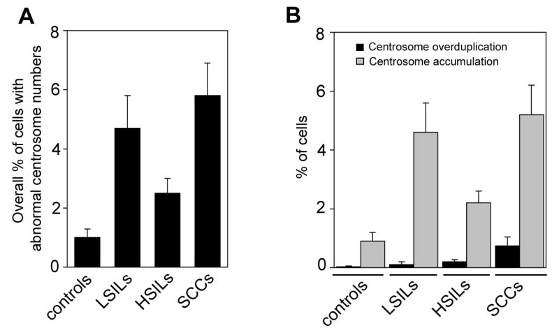
Figure 2. Numerical centrosome aberrations and centrosome overduplication in benign and malignant anal lesions.

(A,B) Quantification of the overall percentage of cells with abnormal centrosome numbers (more than two per cell, A) or centrosome overduplication (more than two per cell with only one centrosome showing positivity for Cep170, B) in hemorrhoids (controls), LSILs, HSILs or anal SCCs. Each bar represents mean and standard error of results from at least 100 cells from three different areas of each sample.
