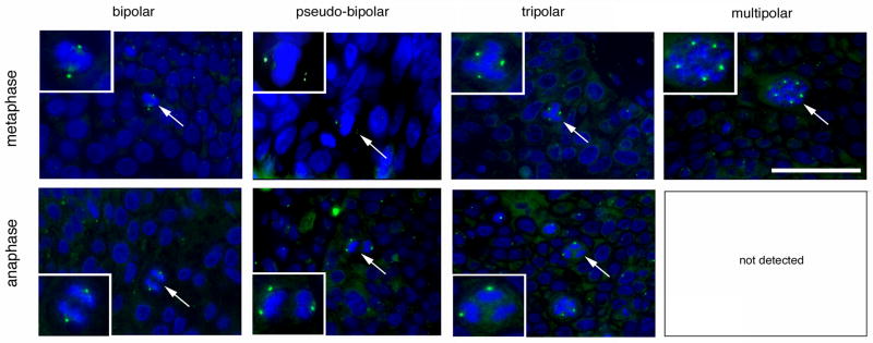
Figure 3. Cell division errors in high-risk HPV-associated anal lesions.
Examples of normal cell divisions and cell division errors during metaphase (top panels) or ana-/telophase (bottom panels) detected by immunofluorescence microscopy for γ-tubulin. The mitotic figures shown in the inserts are highlighted by arrows. Note that multipolar ana-/telophases were not detected in any sample. Nuclei stained with DAPI. Scale bar indicates 50 μm.

