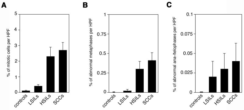
Figure 4. Cell division errors increase with malignant grade in HPV-associated anal lesions.
(A–C) Quantification of overall mitotic cells (A), cells with cell division errors during metaphase (B) or cells with cell division errors during ana-/telophase (C) in hemorrhoids (controls), LSILs, HSILs or anal squamous cell carcinomas (SCCs) per high-power field (HPF). Cell division errors included pseudo-bipolar, tripolar or multipolar configurations. Each bar represents mean and standard error of results from at least 10 HPFs from different areas of each sample.

