
Figure 2. Activation of IRE-1, PERK and ATF6 arms of the UPR in S63del nerves.
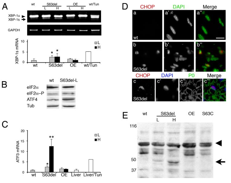
A) IRE-1 catalyzes excision of a 26 nt fragment from XBP-1 mRNA. Semi-quantitative RT-PCR for XBP-1 unspliced (XBP-1u), spliced (XBP-1s), normalized to GAPDH, shows that the spliced XBP-1 is enriched in S63del and tunicamycin-treated nerves. P0wt overexpressor (OE) nerves are included as expression-matched controls for S63del low (L) and high (H). Error bars, s.e.m. for n = 6 individual nerves, *p < 0.05 relative to wt by Student's t-test. B) Levels of eIF2α, eIF2α–P (phosphorylated), and ATF4, were measured by Western analysis. Tubulin (Tub) shows equal loading. C) ATF3 mRNA was measured by quantitative RT-PCR and normalized to phosphoglycerate kinase 1(PGK1) mRNA. ATF3 is induced in a dose-dependent fashion in S63del mice and in tunicamycin-treated livers. Error bars, s.e.m., n = 6, 5, 5, 3, and 3 nerves for wt, S63del-L, S63del-H, OE-L, OE-H, respectively and 3 livers. ** p < 0.01, * p < 0.05 relative to wt by Student's t-test. D) Immunostaining reveals CHOP in elongated, DAPI positive, Schwann cell nuclei in longitudinal sections of S63del nerves at P28 (b–b"), but is undetectable in normal nerves (a–a") Many CHOP-positive Schwann cells contain integral myelin sheaths (c–c"'). Bar = 15 μm in a–b"; 13 μm in c–c"'. E) Western analysis for ATF6 reveals dose-dependent cleavage of the 90kD precursor protein (arrowhead) to the 50kD product (arrow) only in S63del, but not S63C, P0 overexpressor (OE) or wt nerves. Numbers indicate relative molecular weights (Mr).