
Figure 4.
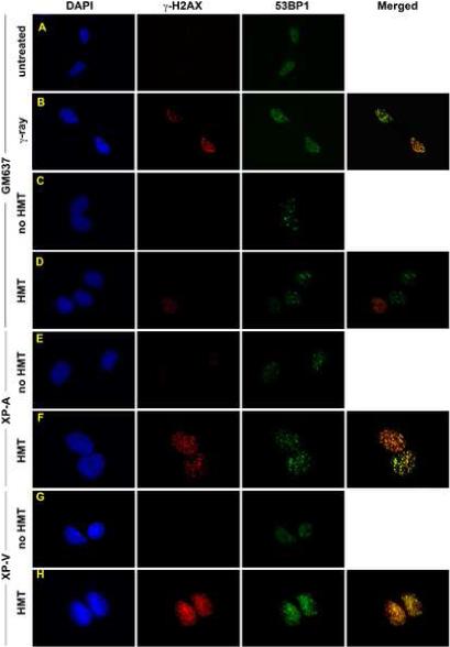
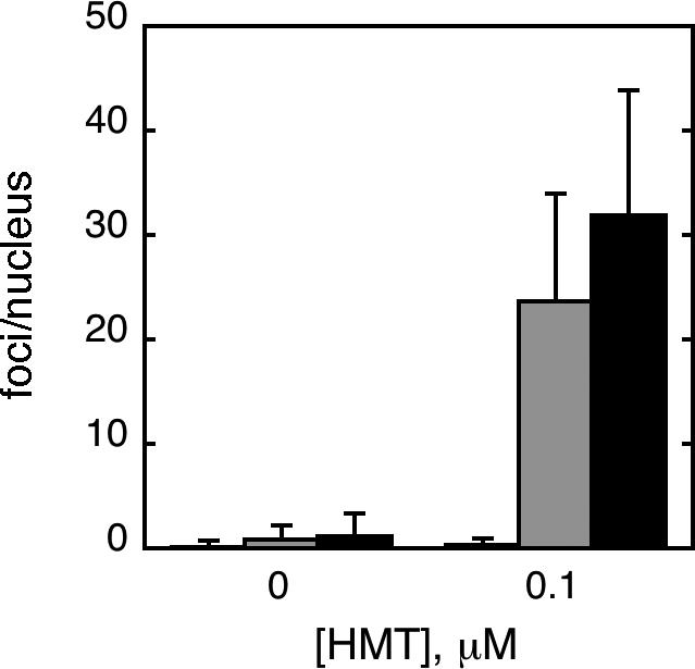
Immunofluorescence of γ-H2AX following HMT and UVA. GM637 cells that were A) untreated, or B) irradiated with 6 Gy of γ-rays and fixed within 15 minutes. GM637 (C, D), XP12RO (E,F), and XP30RO (G,H) cells were incubated either with no HMT (C, E, G) or with 0.1 μM HMT followed by split dose UVA. Cells were allowed to incubate in medium for 6 hours before fixation. All cells were stained with antibodies to γ-H2AX and 53BP1, counterstained with DAPI, and epifluorescence was imaged with a 100× objective. In merged images, yellow color represents sites of coincident γ-H2AX and 53BP1 antibody staining. I) γ-H2AX foci were quantified in nuclei of one hundred randomly selected GM637 (white bars), XP-A (gray bars) and XP-V (black bars) cells treated with or without HMT followed by split-dose UVA to determine the average number of foci per nucleus. Error bars denote standard deviations.