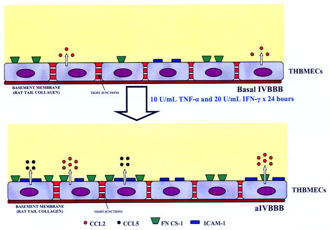
Figure 5.

Functional characteristics of the basal IVBBB and aIVBBB. This figure is a schematic illustration of the effects of cytokine-activation on the IVBBB model. Activating subconfluent THBMECs with 10 U/mL TNF-α and 20 U/mL IFN-γ for 24 hours resulted in increased CCL2 secretion from ~600 pg/mL in the basal IVBBB to ~1200 pg/mL in the aIVBBB. There was no detectable CCL5 secretion by the basal IVBBB, increasing to ~900 pg/mL by the aIVBBB. Cytokine activation induced the expression of ICAM-1 (~20% on the basal IVBBB to >90% on the aIVBBB) and FN CS-1 (~50% on the basal IVBBB to >90% on the aIVBBB) on confluent THBMEC layers in vitro. Other studies demonstrate that the physical and biochemical properties of the IVBBB are not affected by these concentrations of cytokines [40]. Key: aIVBBB: cytokine-activated in vitro blood-brain barrier, FN CS-1: fibronectin connecting segment-1, ICAM-1: intercellular adhesion molecule-1, IFN-γ: interferon-γ, IVBBB: in vitro blood-brain barrier, THBMECs: SV-40 T-antigen immortalized human brain microvascular endothelial cells, TNF-α: tissue necrosis factor-α.
