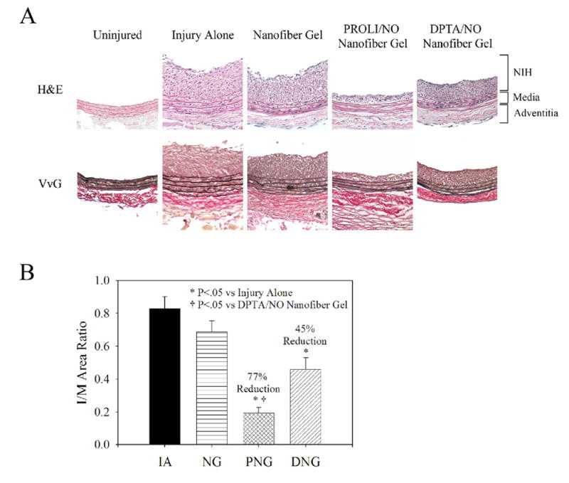
Figure 4.

Rat carotid artery sections from uninjured, injury alone, nanofiber gel, PROLI/NO nanofiber gel, and DPTA/NO nanofiber gel animals sacrificed at 14 days (n=6 per group). A) Representative sections (100x magnification) from each group using routine Hematoxylin and Eosin (H&E) stain and Verhoff-van Gieson (VvG) stain. Graphical representation of B) Intima/Media (I/M) area ratio. Morphometric analysis conducted on 6 sections per rat. Units are arbitrary. IA=injury alone, NG=nanofiber gel, PNG=PROLI/NO nanofiber gel, and DNG=DPTA/NO nanofiber gel.
