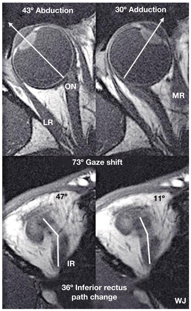
Fig. 1.

Axial MRI images of a right orbit taken at the level of the lens, fovea, and optic nerve (top row), and simultaneously in an inferior plane along the IR muscle path (bottom row), in abduction (left) and adduction (right). Note the bisegmental IR path, with an inflection corresponding to the IR pulley. For this 73° horizontal gaze shift, there was a corresponding 36° shift in IR muscle path anterior to the inflection at its pulley. By permission from Demer [19].
