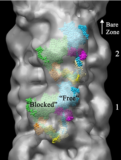
Fig. 2.
Fitting of atomic model of myosin heads in off-state conformation (24) to head motif in crowns 1 and 2 of reconstruction. The 3D envelope has been made translucent to aid visualization of enclosed atomic model. Small regions of atomic model outside the envelope appear bright, whereas the majority, enclosed within it, appears dull (see also SI Movie 2). The heads are labeled “blocked” and “free” according to terminology proposed to describe the actin-binding capability of each head for regulated myosin (24, 25). “Blocked” head: motor domain, green; essential light chain, orange; regulatory light chain, yellow. “Free” head: motor domain, cyan; essential light chain, pink; regulatory light chain, beige. Bare zone direction toward top.

