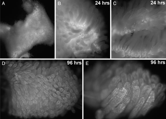
Fig. 5.
Exploring cell proliferation and tissue dynamics in animals using EdU. Whole-mount fluorescent images of mouse small intestine, stained to detect EdU incorporation. Two adult mice were each injected i.p. with 100 μg of EdU in PBS and their small intestines were removed after 24 and 96 h, respectively. A freshly harvested 2-cm-long segment of the small intestine (A) was opened with a longitudinal cut, rinsed, and immediately stained for 10 min with 100 μM TMR-azide in the presence of Cu(I), without fixation or permeabilization. The intestine piece was then fixed, washed to remove unreacted TMR-azide, and imaged at low magnification on a dissecting microscope equipped with fluorescence. (B and C) After 24 h, the EdU shows strong incorporation in cells located at the base of each villus. (D and E) Ninety-six hours after the EdU pulse, the labeled cells have moved away from the base, near the tips of the villi. See text and Materials and Methods for details.

