Fig. 4.
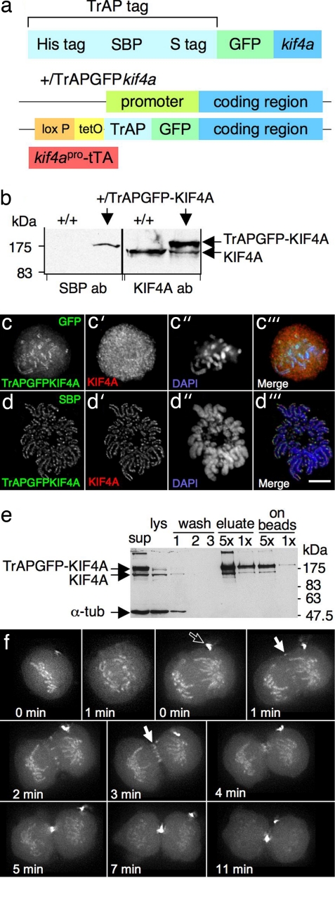
Integration of TrAPGFP in the kif4a genomic locus and application of the cell line. (a) Schematic presentation of TrAPGFPkif4a. (b) TrAPGFP-KIF4A is expressed at similar to wild-type cell level. (c–c‴) Four-percent paraformaldehyde-fixed cells. TrAPGFP-KIF4A localizes axially on chromosomes and diffusely in cytosol during prometaphase. Note that KIF4A-specific antibodies did not show specific staining in prometaphase cells under these fixation conditions. DNA was stained with DAPI. (d–d‴) Cold methanol/acetic acid-fixed cells. TrAPGFP-KIF4A colocalized with endogenous KIF4A on chromosome axes. Note that TrAPGFP-KIF4A was stained by using anti-SBP antibody because the GFP signal was lost after this fixation. (Scale bar: 5 μm.) (e) One-step crude purification of TrAPGFP-KIF4A from lysates of cycling cells with streptavidin beads revealed that TrAPGFP-KIF4A protein is complexed with endogenous KIF4A protein. Equivalent amounts of cells were loaded in each lane unless otherwise indicated. (f) Stills from SI Movie 1of mitosis in a TrAPGFP-KIF4A integrant cell line. TrAPGFP-KIF4A protein localizes on chromosome axes and diffusely in cytosol throughout mitosis. During anaphase, part of KIF4A transfers to the central spindle (filled arrow) and later concentrates at the midzone and midbody. A vestigial midbody from a previous division is labeled with the empty arrow. Because two movies following the same cell continuously were fused, two “time = 0 points” are shown. The first, from the beginning to the onset of anaphase, shows one focal plane. The second, from the onset of anaphase to late cytokinesis, is a projection of 10 focal planes. Time interval, 1 min.

