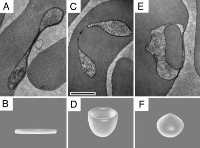
Fig. 1.
Hemoglobin uptake by the Big Gulp. (A, C, and E) TEM images showing the Big Gulp, in which the parasite forms a single large vacuole filled with hemoglobin. (B, D, and F) Illustrations showing the shape of the ring-stage parasite throughout the Big Gulp process. (A and B) The ring-stage parasite as it appears shortly after red blood cell invasion when it has flattened and taken on a biconcave disk shape. The parasite then takes on the shape of a cup (C and D). (E and F) Completion of the Big Gulp when the parasite has taken a single large vacuole filled with hemoglobin. (Scale bar, 0.5 μm.) Fig. 2 shows that this vacuole can be as large as 40% of the parasite volume. SI Movies 3–6show four full serial sectioned rings at different stages of this process.

