Fig. 5.
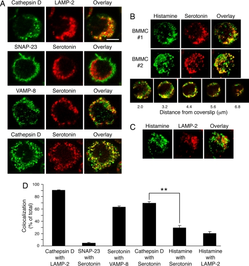
Histamine and serotonin are present in distinct secretory granule subsets in mast cells. Mast cells isolated from wild-type mice were stained with the indicated antibodies and analyzed by confocal immunofluorescence microscopy. (A) Single 0.4-μm-thick optical sections of representative images are shown. (Scale bar: ≈6 μm.) (B) Three-dimensional reconstruction of serial sections from two representative mast cells stained with histamine (green) and serotonin (red) antibodies and an overlay (yellow) are shown. An overlay of individual serial sections of a mast cell stained with histamine (green) and serotonin (red) antibodies reveals heterogeneity within the secretory granule population. (C) Three-dimensional reconstruction of serial sections from a representative mast cell stained with histamine (green) and LAMP-2 (red) antibodies and an overlay (yellow) are shown. (D) The percentage of colocalization was calculated for each combination of markers by voxel analysis of each confocal plane of >20 cells. There was a statistically significant difference in the colocalization of cathepsin D with serotonin as compared with colocalization of histamine with serotonin (**, P < 0.001).

