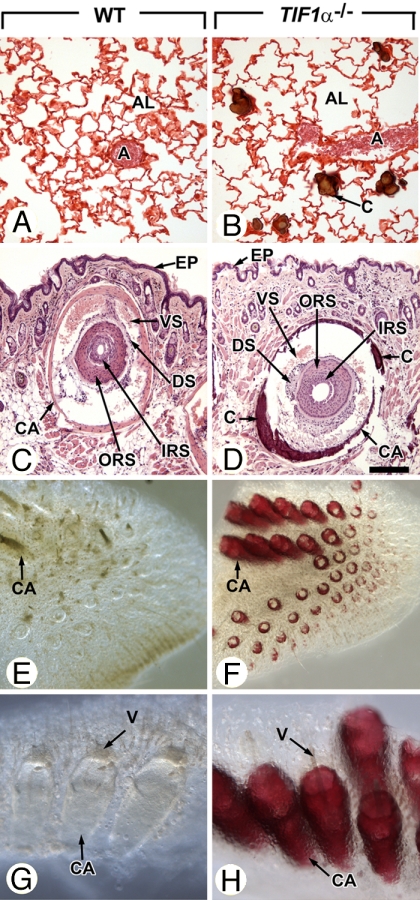
Fig. 2.
Ectopic calcifications of lungs and vibrissae in 10-month-old TIF1α−/− mutants. (A–D) Histological sections through lungs and vibrissae of TIF1α−/− mutants and age-matched WT littermates stained by using the von Kossa method (brown calcified deposits) and either safranin 0 (A and B) or hematoxylin/eosin (C and D). (A and B) Note that calcified nodules are restricted to the TIF1α−/− mutant alveoli, are not associated with inflammatory cell infiltrates, and spare the lung arterial bed. (C and D) Transverse sections through vibrissae surrounded by blood vessel sinuses (VS), which are themselves encased by a muscular capsule (CA). (E–H) Low (E and F) and high (G and H) high- magnification views of whole-mounts of muzzle skin stained with alizarin red confirm extensive mineralization of vibrissae capsules in TIF1α−/− mutants. Note that photomicrographs of whole-mounts in E and F and in G and H were taken at identical magnifications. A, arteries; AL, lung alveoli; C, calcified deposits; CA, capsules of vibrissae; DS, ORS and IRS, dermal sheath, outer root sheath and inner root sheath of the vibrissae, respectively; EP, epidermis; V, vibrissae; VS, blood vessel sinuses of the vibrissae. (Scale bar: A and B, 80 μm; C and D, 160 μm.) (Magnifications: E and F, ×10; G and H, ×60.)

